Getting a closer look inside biomolecules
Novel isotope tracking brings nanoscale chemistry into view

The Science
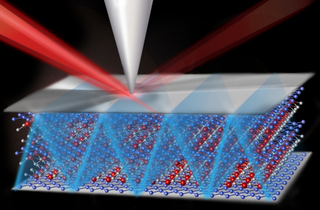
Isotope “labeling” techniques replace specific atoms in a compound with an isotope that can be detected by its neutron count. For example, it may replace naturally-occurring carbon-12 with carbon-13. Tracking labeled isotopes can help researchers see what happens during complex chemical reactions inside cells or other organic samples. Researchers can then better understand the roles of proteins and amino acids in many cellular processes. Conventional techniques to detect labels can destroy the samples. In addition, these techniques’ resolution is limited. A new method using electron microscopy expands the options that scientists can use to study sensitive samples without destroying them. It also offers better resolution than conventional methods.
The Impact
Using electron microscopy to track isotopes in organic samples can provide a major new tool for chemistry and biology research. This technique paves the way for new experiments that study complex biochemical reactions in samples that contain entire cells. In particular, it could enable scientists to study these samples at much smaller sizes than they can now, down to the nanoscale.
Summary
Isotope labeling is widely used to obtain detailed structural information about biomolecules such as proteins or amino acids. Several methods can be used to track isotopes by their mass or vibrational modes, but most reliable methods are destructive and can change or damage samples. Additionally, conventional methods are limited to macroscale resolution and require large sample sizes that may be difficult to produce in biological experiments.
To overcome these challenges, researchers demonstrated the first use of electron microscopy to identify site-specific isotopic labels at nanoscale resolution. Vibrations between atoms in the amino acid L-Alanine were measured without damaging the sample using monochromated electron energy loss spectroscopy (EELS).
Contact
Jordan Hachtel
Center for Nanophase Materials Sciences, Oak Ridge National Laboratory
hachtelja@ornl.gov
Funding
The research was funded by the Department of Energy, Office of Science, Office of Basic Energy Sciences, Chemical Sciences, Geosciences, and Biosciences Divisions. It used resources of the Center for Nanophase Materials Sciences and the National Energy Research Scientific Computing Center, which are both DOE Office of Science User Facilities.
Publications
J.A. Hachtel, et al., “Identification of site-specific isotopic labels by vibrational spectroscopy in the electron microscope.” Science 363, 525–528 (2019). [DOI: 10.1126/science.aav5845]
Related Links
Oak Ridge National Laboratory press release: Novel electron microscopy offers nanoscale, damage-free tracking of isotopes in amino acids
Highlight Categories
Performer: DOE Laboratory , Industry , SC User Facilities , ASCR User Facilities , NERSC , BES User Facilities , CNMS